In summary these findings are consistent with a normal examination of the spine For completeness I would like to. The iliac crest is the part of the ilium that stretches between the posterior superior and anterior superior iliac spine.

Which Spinal Levels Are Identified By Palpation Of The Iliac Crests And The Posterior Superior Iliac Spines Chakraverty 2007 Journal Of Anatomy Wiley Online Library
Now work your way down to the ILAs and determine if there is a posteriorinferior ILA.

. The abdominal wall is split into the posterior back lateral sides and anterior front walls. The pelvis must be stabilized. It lies deep in the popliteal fossa and requires deep palpation to feel.
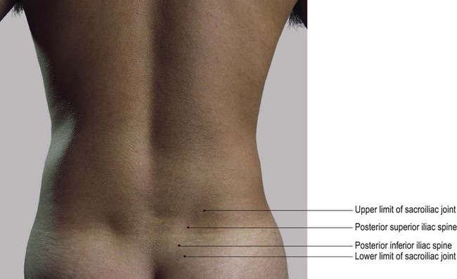
50 Patients with any of these conditions can experience pain near the posterior superior iliac spine and may have buttock pain that radiates down the leg. Identify the location of the posterior superior iliac spine PSIS on each side. The sartorius and tensor fasciae latae muscles of the thigh begin at the anterior superior iliac spine.
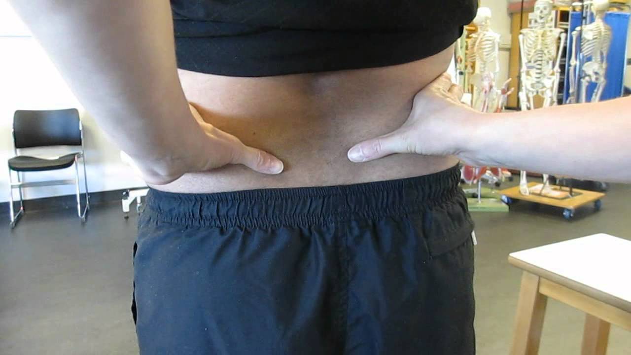
Begin by finding the PSISs by following the iliac crests posteriorly. To make it easier you can ask the. The two vertical or mid-Poupart lines are drawn from the point midway between the anterior superior spine and the pubic symphysis on each side vertically upward to the costal margin.
Posterior branch This supplies. Place pressure on the sulci and determine if there is a deep sulcus. The patient lies supine and the examiner flexes abducts and rotates the patients affected joint.
The range of movement of the cervical thoracic and lumbar spine was normal. The right one is the most valuable as the ileo-caecal valve is situated where it cuts the. From the PSISs move slightly medial and superior about 1cm so that you are now in the sacral sulci.
This technique requires training in bony-landmark identification. If the joint side that is flexed moves up this is considered a positive test. The transverse abdominis muscle and the.
The femoral pulse can be palpated as it enters the femoral triangle midway between the anterior superior iliac spine of the pelvis and the pubis symphysis the mid-inguinal point. Next perform the spring test by first finding the. The examiners thumbs are placed under the posterior superior iliac spine and S2.
SI joint dysfunction has a variety of causesincluding hypermobility hypomobility trauma degenerative arthritis inflammatory arthropathy sacroiliitis infection ligament strain andor stress fractures. The patient is asked to stand on one leg while moving the opposite leg towards the chest. Many different muscles of the thigh and trunk are connected to the iliac crest.
Assessment of the spine revealed normal alignment with no tenderness on palpation. The popliteal artery is the hardest pulse to find. Pathology of the SI.
However the anterior tilt may require a more closely monitored palpation technique always get your clients permission before touching where you identify the location of the anterior superior iliac spine and compare it to the position of the posterior superior iliac spine to see if there is an excessive tilt.

Finding Anterior And Posterior Iliac Spines Superior And Inferior Youtube

Psis Palpation At Superior Aspect Download Scientific Diagram

Muscle And Bone Palpation Of The Low Back And Pelvis
Surface Anatomy Pelvis Posterior Landmarks On Model Youtube

The Reliability Of Palpating The Posterior Superior Iliac Spine A Systematic Review Semantic Scholar
The Abdomen Basicmedical Key

Palpation Of Posterior Superior Iliac Spine Download Scientific Diagram

Posterior Superior Iliac Spine Wikipedia
0 comments
Post a Comment